
Reading Time:
Overview
In a radiologic exam, a penetrating beam of X-rays transilluminates the patient, showing tissues of differing densities of mass within the body as images of differing intensities (areas of relative light and dark) on the film or monitor.
A tissue or organ that is relatively dense in mass (e.g. bone) absorbs or reflects more X-rays than a less dense tissue (e.g. the spleen). Consequently, a dense tissue or organ produces a brighter (more white-looking) area on a monitor as fewer X-rays reach the film or detector. A dense substance is radiopaque, whereas a substance of less density is radiolucent.
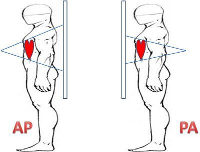
In radiologic nomenclature, postero-anterior (PA) projection refers to a radiograph in which the X-rays pass-through the patient from posterior (P) to anterior (A) i.e. the X-ray tube was posterior to the patient and the X-ray film or detector was anterior. A radiograph using anteroposterior (AP) projection radiography is the opposite.
The introduction of contrast media (radiopaque fluids such as iodine compounds or barium) allows the study of various luminal or vascular organs and potential or actual spaces (such as the digestive tract, blood vessels, kidneys, synovial cavities, and the subarachnoid space) that are not visible on plain films.
Key Images
Key Info
Radiographs are also sometimes referred to as X-Rays or plain films. Structures that are dense reflect the x-ray beam and appear light/white on the monitor e.g. bones. The standard chest radiograph (CXR) is taken in the PA position to minimise enlargement artefact of the heart (think of it like an object casting a shadow producing a larger shadow the nearer the object is to the source of the light it obstructs).
Clinical Anatomy
Standard radiographs include chest, abdomen, pelvis and films of specific limbs when looking for fractures.
Radiographs should also ways be taken from two angles as they are 2D rather than 3D and changes may be missed if only looking at one dimension. Two views also help to localise any abnormalities more accurately.
Summary
Structures that are dense reflect the x-ray beam and appear light/white on the monitor e.g. bones.