Reading Time:
Overview
Computed tomography or CT scans show radiographic images of the body that resemble transverse anatomical sections. In this technique, a beam of X-rays passes through the body as the X-ray tube and detector rotate around the axis of the body. Multiple overlapping radial energy absorptions are measured, recorded, and compared by a computer to determine the radiodensity of each volumetric pixel (voxel) of the chosen body plane. The radiodensity of (amount of radiation absorbed by) each voxel is determined by factors that include the amount of air, water, fat, or bone in that element.
The computer maps the voxels into a planar image (CT slice) that is displayed on a monitor.
Depending on the structure being imaged, CT scans can be used with and/or without contrast.
Key Images

Key Info
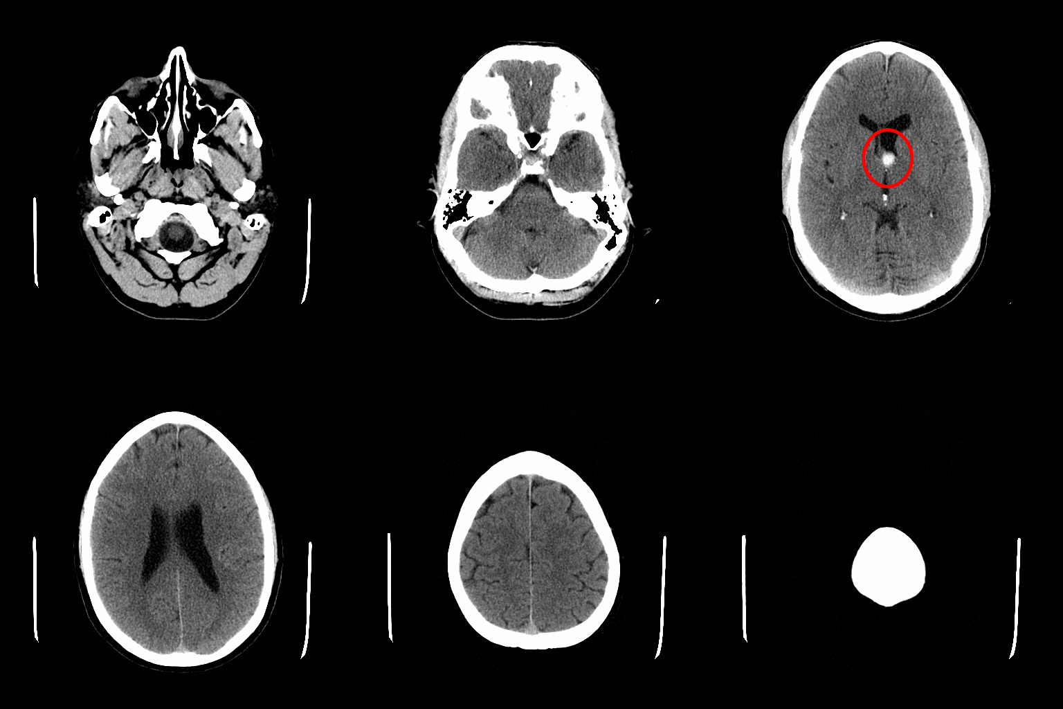
CT images relate well to conventional radiographs, in that areas of great absorption (e.g., bone) are relatively transparent (white) and those with little absorption are black. CT scans are always displayed as if the viewer were standing at a supine patient’s feet—i.e., from an inferior view.
When interpreting at CT scan, it is important to determine the orientation. Images are most commonly presented in the transverse plane, and are orientated so that we are looking up the body from the patient’s toes.
A helpful way to get your bearings is the acronym RALP. Starting at the 9 o’clock position and moving clockwise in 90 degree intervals, we are looking at the Right, Anterior, Left and Posterior aspects of the patient.
Clinical Anatomy
The introduction of an intravenous radiofluorescent contrast into the bloodstream can be used for a variety of diagnostic purposes, for example:
- Used to visualise the cardiovascular system (e.g. investigating for suspected aneurysms, dissections, or atherosclerotic diseases).
- Used to identify whether a tumour is malignant.
After approximately 7 minutes following an intravenous injection with iodinated CT contrast, the contrast begins to expel from the body via the urinary system. The contrast can be seen in the ureters going into the bladder creating a CT Urogram; a procedure that is commonly replacing the traditional intravenous pyelogram seen in radiography.
Oral contrast can also be administered if investigation is required of the digestive system. (Crohn’s disease, bowel obstruction, diverticulitis, appendicits).
Intracranial bleeds are potentially life-threatening conditions, and occur most commonly as an acute or delayed response to trauma. They can occur spontaneously from the rupture of cerebral aneurysms, but this is less common.
CT scanning has evolved to become to mainstay of investigation of patients with a suspected intracranial bleed.
Summary
CT scans are created using a series of x-rays, which are a form of radiation on the electromagnetic spectrum. The scanner emits x-rays towards the patient from a variety of angles – and the detectors in the scanner measure the difference between the x-rays that are absorbed by the body, and x-rays that are transmitted through the body. This is called attenuation.
The amount of attenuation is determined by the density of the imaged tissue, and they are individually assigned a Hounsfield Unit or CT Number.
High density tissue (such as bone) absorbs the radiation to a greater degree, and a reduced amount is detected by the scanner on the opposite side of the body
Low density tissue (such as the lungs), absorbs the radiation to a lesser degree, and there is a greater signal detected by the scanner.
References
RL Drake, W Vogl, AWM Mitchell. Gray's Anatomy for Student (2005). 39th Ed. Ediburgh: Elsevier.
