Reading Time:

Overview


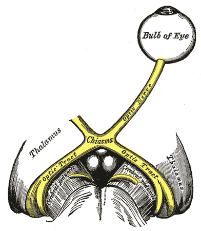
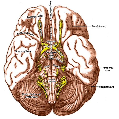
The optic nerve (CNII) is the nerve of sight. It is not a true cranial nerve as it develops as an outpouching of the diencephalon (part of the brain itself). The two optic nerves merge at the chiasm and separate again as the optic tracts. These tracts then run to the lateral geniculate nucleus of the thalamus, and then to the visual loops and then to the primary visual cortex in the depths of the calcarine sulcus of the occipital lobe.

Gross Anatomy


The optic nerves develop from outpouchings of the diencephalon (part of the brain itself) and are hence part of the central not peripheral nervous system. This is reflected in the myelin that coats the nerves, which is composed of oligodendrocytes, and not schwann cells like the rest of the peripheral nervous system. The optic nerve is covered by the meningeal layers, rather than the epineurium, perineurium, and endoneurium that cover the other cranial nerves.

It receives its sensory information from the retinal cells (rod cells for black and white, and cone cells for colour) that line the retinal epithelium, located in the eye. This includes light, colour, brightness, visual acuity, and contrast. The central part of the retina i.e. the central vision of the eye is an area densely packed with retinal cells, and has a high degree of acuity. This is the fovea centralis.

These retinal cells transmit the visual information they receive to photosensitive ganglion cells, which all ultimately converge onto the optic nerve, which then travels posteromedially through the optic canal. Where the optic nerve leaves the back of the eye, there is an absence of retinal cells. This is the blind spot and is enlarged in raised intraocular pressure. The nerve then unites with its counterpart from the opposite side at the optic chiasm (so named due to its resemblance to the Greek letter chi). An important thing to realise is that everything in the eye crosses over. The light from your temporal visual field, strikes your nasal (medial) retina. The light from your nasal field strikes your temporal retina. 

At the chiasm, the fibers from your the temporal visual field (nasal retina) cross to the other side. The nasal visual field (temporal retina) does not cross over). The optic chiasm then sends back two optic tracts, that synapse in the lateral geniculate nucleus of the thalamus (the thalamus is the gateway to the cerebral cortex, and all sensory information apart from olfactory passes through it). The optic nerve also sends projections to the superior colliculi and suprachiasmatic nucleus (our internal light sensitive clock).

This then is sent back through the optic radiation through Meyer’s loop (temporal loop, superior visual field, inferior retina) and Baum’s loop (parietal lobe, inferior visual field, superior retina), in order to reach the primary visual cortex (located in the occipital lobe, within the depths of the calcarine sulcus). Secondary visual cortices are located close by and interpret the visual information we see in further detail.

The optic nerve mediates the accommodation and light reflexed. The accommodation reflex is when the lens of the eye relaxes and becomes swollen and round when looking at a near object. The light reflex refers to constriction of both pupils of the eye in response to light.

Clinical Anatomy


Homonymous hemianopia- A stroke occurring within the optic tract (post chiasm projection to the lateral geniculate nucleus of the thalamus), results in a homonymous hemianopia. The nasal visual field on the affected side, and the temporal retinal shield on the unaffected side are affected. The patient will complain of frequently bumping into things.


Glaucoma- This is a condition characterised by progressive visual loss and optic nerve damage. A rise in intraocular pressure is a major risk factor (possibly sue to decreased drainage of the aqueous humor in the anterior chamber of the eye). The condition is divided into closed angle and open angle. Open angle is painless, and develops chronically over time. Closed angle is also chronic, but is usually painful and may present acutely. The patient may also complain of sudden blurred vision, nausea and vomiting.


Pituitary Tumour- The pituitary gland sits in the sella turcica, a bony cavity within the body of the sphenoid bone. This cavity lies directly beneath the optic chiasm. If the pituitary gland enlarges e.g. due to a tumour, the tumours may compress the chiasm from beneath. This affects the fibers that cross over (from the temporal visual fields, which corresponds to the nasal retina), and results in bitemporal hemianopia. Patients lose their peripheral vision, which results in the patient knocking into things as they walk or drive.


Diabetic retinopathy- Glucose in the micro-arteries of the retina is very damaging. Changes to the retina occur, following from the ischaemia. Cotton wool spots, flame haemorrhages, exudates and aneurysms result. In advanced disease, neovascularisation begins, and may affect the macula, compromising the patient’s central high quality vision.


Quick Anatomy


Key Facts

Developmental Precursor: Diencephalon (secondary brain vesicle)
Special Sensory: Sight

Aide-Memoire

Every cranial nerve related to the eye apart from the optic nerve, (CNII) leaves the skull cavity through the superior orbital fissure:
Oculomotor (CNIII), 
Trochlear (CNIV), 
Ophthalmic division of the trigeminal (V1)
Abducens (CNVI) 

Summary


The optic nerve (CNII) is the nerve of sight. The two optic nerves merge at the chiasm and separate again as the optic tracts. 

References


Frank H.Netter MD: Atlas of Human Anatomy, 5th Edition, Elsevier Saunders, Chapter 1 Head and Neck
Chummy S.Sinnatamby: Last’s Anatomy Regional and Applied, 12th Edition, Churchill Livingstone Elsevier
Richard L. Drake, A. Wayne Vogl, Adam. W.M. Mitchell: Gray’s Anatomy for Students, 2nd Edition, Churchill Livingstone Elsevier
Elliiot L.Manchell: Gray's Clinical Neuroanatomy: The Anatomic Basis for Clinical Neuroscience
The Definitive Neurological Surgery Board Review By Shawn P. Moore, 2005
Human Neuroanatomy By James R. Augustine, 2008